

Welcome to the BEATS (BEAmline for Tomography at SESAME) beamline.
The BEATS beamline is located at port I10 of the SESAME storage ring and operates a hard X-ray full-field radiography and micro Computed Tomography (CT) station. Hard x-ray imaging can be applied in numerous scientific areas.
The layout of BEATS is inspired by TOMCAT, the tomography beamline at the Swiss Light Source (SLS). BEATS is optimized for absorption and phase contrast X-ray radiography and tomography.
The BEATS beamline was constructed with generous funding from the European Union’s Horizon 2020 programme. It was built and commissioned by a consortium of leading research facilities in the Middle East (SESAME and The Cyprus Institute), well-established European synchrotron radiation facilities and high-energy laboratories with decades-long experience in synchrotron radiation research and technology DESY (Germany), Elettra (Italy), ESRF (France), INFN (Italy) and PSI (Switzerland), and more recently-founded synchrotron laboratories ALBA-CELLS (Spain) and SOLARIS (Poland).
BEATS has been opened to users in the second 2023 call for proposals for which the deadline for the submission of applications is 30 September 2023.
The layout of BEATS is inspired by TOMCAT, the tomography beamline of the Swiss Light Source (SLS) [1]. The total length of the BEATS beamline is 45.3 m. The insertion device is a 2.9 T 3-Pole Wiggler (Ecritical = 12 kEv) that provides photon energies well above 80 keV. The beamline can be operated in two modalities: filtered white beam, and monochromatic beam using a Double Multilayer Monochromator (DMM).
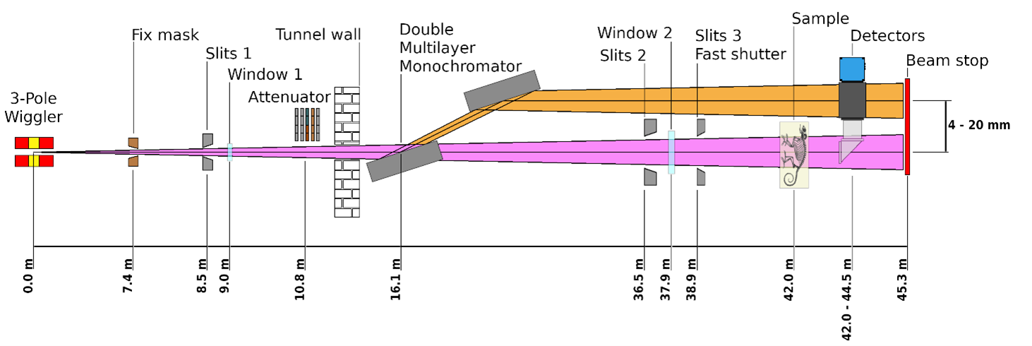
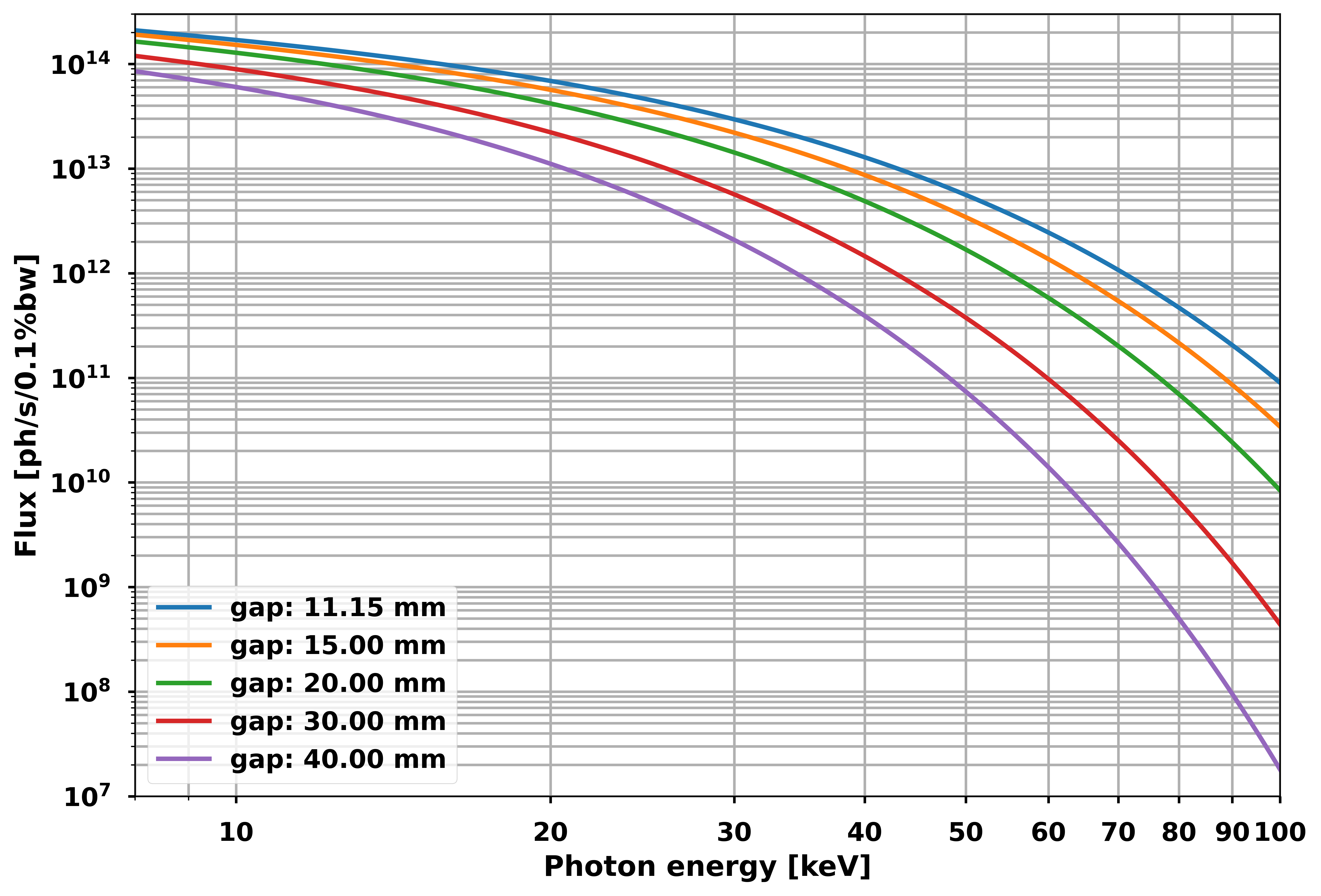
The usable beam size at the sample position is 70 x 15 mm2, with estimated flux as high as 1×10¹⁰ Ph/s/mm2 in 0.1% of the source bandwidth (white beam configuration).
X-ray source
The BEATS photon source is a 3-pole wiggler of 2.9 T peak magnetic field, operating in a SESAME short straight section at a variable gap. The design of the insertion device is compact and gives low multipolar effects on the SESAME storage ring optics, while as well reducing the attractive forces between the magnetic structures leading to minor mechanical constraints [2]. The critical energy of the X-ray source is 12 keV, and the energy spectrum reaches 100 keV.
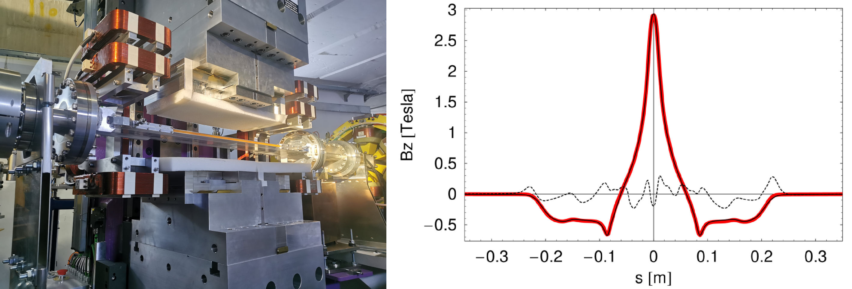
| BEATS 3-POLE WIGGLER PARAMETERS | |
|---|---|
| Type | 3-pole wiggler |
| Minimum gap | 11.15 mm |
| Peak field | 2.9 T |
| Critical energy | 12.0 keV |
| Magnetic length | 0.41 m |
| Manufacturer | Kyma S.p.A. (Italy) |
Front-end
The BEATS front-end was designed taking the standard configuration for bending magnet Front-Ends at ALBA, adapting it to the particularities of SESAME [2]. The design includes: (i) a fixed mask to protect the downstream elements and to define the user aperture, (ii) a photon shutter, (iii) a fast closing valve triggered by gauges installed on the beamline side for protection of the accelerator’s vacuum, (iv) a system of primary slits (refurbished from ID19 beamline at ESRF), (v) a set of beam attenuators with 5 independent axes, and (vi) a single-block Bremsstrahlung stopper. All power absorbing elements have been dimensioned to withstand the power delivered by a stored electron beam of up to 400 mA.
Most of the FE units were manufactured by JJ X-ray A/S (Denmark).
Optics
The main optical component of BEATS is a Double Multilayer Monochromator (DMM) placed outside of the SESAME storage ring tunnel in a dedicated Optics Hutch [3]. The DMM allows to select a photon energy between 8 and 50 keV with an energy bandwidth of approximately 2%. 500-mm-long mirrors allow to preserve the usable beam height even when working at high energies. The design is optimized to guarantee stability and minimize vibrations of the two optical elements. The mirrors can be completely removed from the beam path, allowing operation in white-beam mode.
| BEATS MONOCHROMATOR PARAMETERS | |
|---|---|
| Distance from source (1st mirror) | 15.165 [m] |
| Max. Beam size @ 1st mirror | 29 mm × 6 mm (Hor. × Ver.) |
| Offset (variable) | Min. 4.0 – Max. 18.0 [mm] |
| Substrate dimension | 500 mm × 70 mm × 60 mm |
| Distance between ML centers | 510 mm |
| Theta (Bragg angle) | -0.5 – 2.5 [deg] |
| Bragg resolution | 0.5 µrad |
| Energies | 8 – 50 keV |
| Max. power on 1st mirror | 133 W |
| Manufacturer | CINEL Scientific Instruments S.r.l. (Italy) |
| BEATS MULTILAYERS | STRIPE 1 | STRIPE 2 |
|---|---|---|
| Coating | [W/B4C]100 – d = 2.5 nm | [Ru/B4C]65 – d = 4.0 nm |
| d-spacing | 2.5 nm | 4 nm |
| d-spacing gradient along Z | 3.41 % (0.00682 % / mm) | 3.52 % (0.00704 % / mm) |
| N. bilayers | 100 | 65 |
| Energies | 20 – 50 keV | 8 – 22 keV |
| dE/E | 1.8 % | 2.4 % |
| Theta (Bragg angle) | 0.292 – 0.728 [deg] | 0.42 – 1.159 [deg] |
| Filling factor Γ | 0.5 | 0.5 |
| Manufacturer | ESRF Multilayer | Laboratory (France) |
Experimental station
The BEATS experimental hutch covers a length of 9 m. It comprises two sets of beam-defining slits, a CVD diamond window separating vacuum and air sections of the beamline, and the tomography endstation. The sample is mounted at a distance of 43 m from the X-ray photon source.
Endstation #1
The beamline endstation #1 is composed of a sample air-bearing tomography manipulator that was previously at the TOMCAT beamline of SLS [1], and of indirect X-ray detectors installed on a common granite optical table. Object pixel sizes in the range from 13.0 μm down to 0.65 μm are achieved. The usable beam size at the sample position is 70 x 15 mm2.
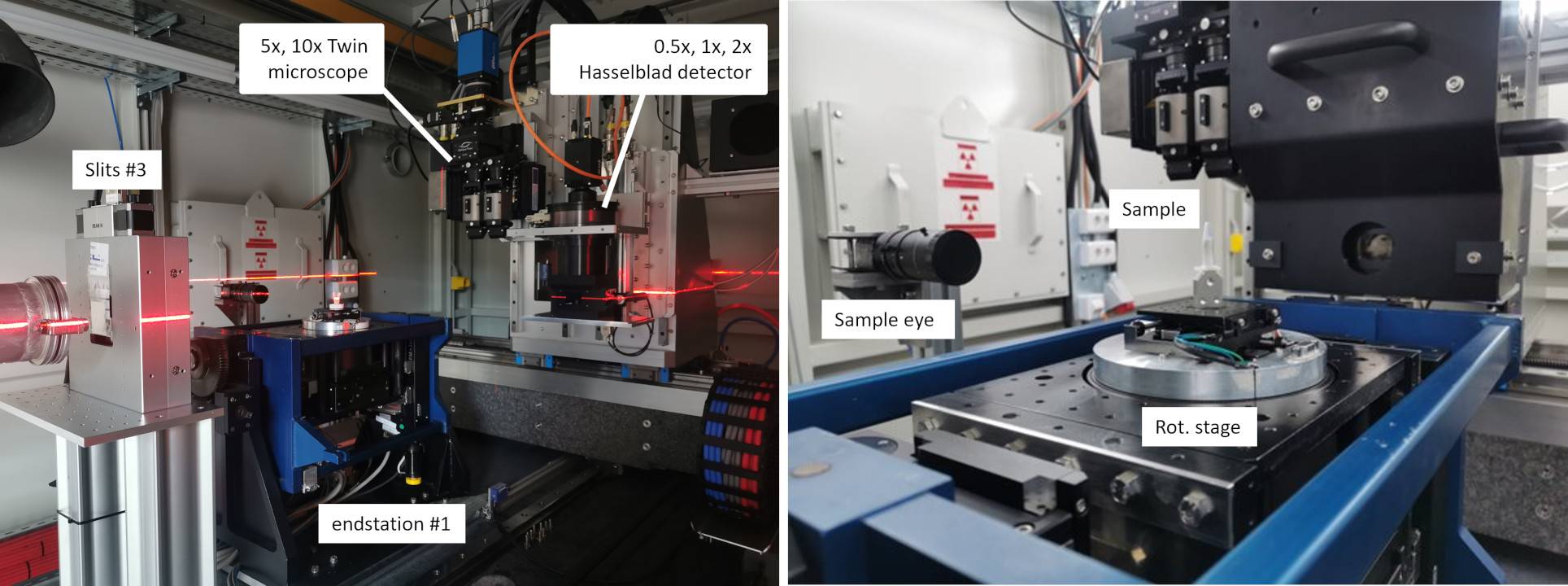
Detectors
| DETECTOR 1 | DETECTOR 2 | DETECTOR 3 | |
|---|---|---|---|
| Type | White-beam medium resolution detector | White-beam Twin microscope | Monochromatic microscope |
| Manufacturer | ESRF (France) | Optique Peter (France) | Optique Peter (France) |
| Lens type | Hasselblad H system | Mitutoyo M Plan Apo(Radiation hardened) | Olympus PLAPO/UPLAPO |
| Magnification | 0.5×; 1×; 2× | 5×; 7.5×; 10× | 10×; 20× |
| Available voxel size (with PCO edge 5.5) |
13 – 3.25 μm | 1.3 – 0.65 μm | 0.65 – 0.33 μm |
Cameras
| CAMERA 1 | CAMERA 2 | |
|---|---|---|
| Sensor type | sCMOS - Mono | CMOS - Mono |
| Manufacturer | PCO (Germany) | Teledyne FLIR (US) |
| Model | edge 5.5 | Oryx 10GigE |
| Megapixels | 5.5 | 7.1 |
| Sensor size | 2560 × 2160 | 3208 × 2200 |
| Pixel size | 6.5 μm | 4.5 μm |
| Max frame rate (full frame) | 100 fps | 112 fps |
| Shutter | Rolling / Global | Global |
| Exposure time | 500 μs - 2 s | 10 μs - 30 s |
| Bit-depth | 16 bit | 8 bit / 16 bit |
| ADC | 16 bit | 8 bit / 10 bit / 12 bit |
| Dynamic range | 88.6 dB | 71.7 dB |
[1] M. Stampanoni et al., “TOMCAT: A beamline for TOmographic Microscopy and Coherent rAdiology experimenTs,” AIP Conference Proceedings, vol. 879, no. 1, pp. 848–851, Jan. 2007, https://doi.org/10.1063/1.2436193.
[2] J. Campmany et al., “Design of Front End and a 3-Pole-Wiggler as a Photon Source for BEATS Beamline at SESAME,” in Proc. IPAC’21, in International Particle Accelerator Conference. Campinas, Brazil: JACoW Publishing, Geneva, Switzerland, Aug. 2021, pp. 324–326. https://doi.org/10.18429/JACoW-IPAC2021-MOPAB086.
[3] G. Iori et al., “Design and Ray-Tracing of the BEATS Beamline of SESAME,” presented at the 11th Mechanical Engineering Design of Synchrotron Radiation Equipment and Instrumentation (MEDSI’20), Chicago, IL, USA, 24-29 July 2021, JACOW Publishing, Geneva, Switzerland, Oct. 2021, pp. 246–248. https://doi.org/10.18429/JACoW-MEDSI2020-WEPA10.
3PW
DMM
Endstation #1 - PSI TOMCAT
MANIPULATOR OR SAMPLE STAGE
Low magnification white-beam detector
DETECTION
White-beam double objective microscope
DETECTION
For information on how to prepare your experiment and analyze your data visit the BEATS User Manual.
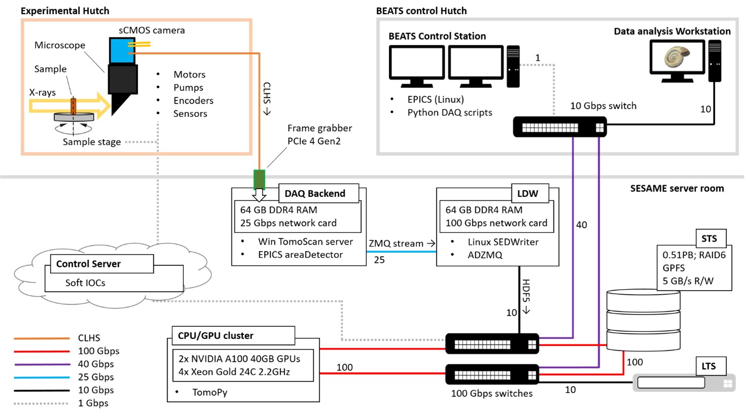
Experiments at BEATS can produce up to 2 Terabytes of experimental data per day, with a peak throughput of 1.1 Gigabyte of data per second. The beamline data acquisition and computing layout follows a network topology and design principles established at the Paul Scherrer Institut (PSI) and European Synchrotron Radiation Facility (ESRF) [1]. All BEATS detector cameras are connected to dedicated backend DAQ servers located in the SESAME server room through Camera Link® HS or 10 Gigabit Ethernet interfaces. The TomoScan Python module is adapted and used as tomography DAQ sub-system. TomoScan is a unified Python module developed at the Advanced Photon Source (APS, Chicago, USA) to collect computed tomography data. It uses PyEpics: the library for EPICS Channel Access for Python developed by the GSECARS team at Argonne National Laboratory and allows to control several cameras and data acquisition systems via EPICS areaDetector. The streaming process of detector data is managed through the asynchronous messaging library ZeroMQ (ZMQ) by the EPICS areaDetector plugin ADZMQ developed at the Swiss Light Source — Paul Scherrer Institut. The ZMQ subscriber was developed at SESAME as part of the SESAME Experimental Data writer (SEDWriter) to handle the creation of HDF5 files according to the Scientific Data Exchange guidelines [2].
Tomographic reconstructions are performed on a dedicated hybrid CPU/GPU cluster. All experimental data is stored on a 0.51 Petabyte GPFS Short Term Storage in compliance with the SESAME Experimental Data Management Policy.
[1] G. Iori, S. Matalgah, C. Chrysostomou, A. Al-Dalleh, and M. Alzu’bi, “Data Acquisition and Analysis at the X-ray Computed Tomography Beamline of SESAME,” in 2021 IEEE Jordan International Joint Conference on Electrical Engineering and Information Technology (JEEIT), Nov. 2021, pp. 134–139. https://doi.org/10.1109/JEEIT53412.2021.9634151.
[2] F. De Carlo, D. G ̈ursoy, F. Marone, et al., “Scientific data exchange: A schema for HDF5-based storage of raw and analyzed data,” J Synchrotron Rad, vol. 21, no. 6, pp. 1224–1230, Nov. 1, 2014, Number: 6 Publisher: International Union of Crystallography, ISSN: 1600-5775. https://doi.org/10.1107/S160057751401604X.
2024 (2), 2023 (2), 2021 (3), 2020 (1), All (8)
2024
- Alrecon: computed tomography reconstruction web application based on Solara
Open Research Europe, Vol. 4 - 54, pp. (2024)
G. Iori, I. Foudeh, M. Alzubi, M. Almohammad, S. Matalgah
doi: 10.12688/openreseurope.16863.1 - Non-destructive 3D exploration of silicate glass corrosion: a combined multiscale approach from the macro to the nanoscale
Physical Chemistry Chemical Physics, Vol. , pp. (2024)
G. Franceschin, R. Zanini, G. Iori, E. Longo, G. Divitini, G. Tromba, A. Traviglia
doi: 10.1039/d3cp05221d
- Ciclope: micro Computed Tomography to Finite Elements
JOSS - Journal of Open Source Software, Vol. 8 - 84, pp. 4952 (2023)
G. Iori, G. Crimi, E. Schileo, F. Taddei, G. Fraterrigo, M. Pani
doi: 10.21105/joss.04952 - An FEA Investigation of the Vibration Response of the BEATS Detector Stage
R&D Journal of the South African Institution of Mechanical Engineering, Vol. 39, pp. 44-52 (2023)
F. Mokoena, M. Bhamjee, S. Connell, P. Van Vaerenbergh, G. Iori, A. Kaprolat
doi: 10.17159/2309-8988/2023/v39a5
- Data Acquisition and Analysis at the X-ray Computed Tomography Beamline of SESAME
2021 IEEE Jordan International Joint Conference on Electrical Engineering and Information Technology (JEEIT), Vol. , pp. 134-139 (2021)
G. Iori, S. Matalgah, C. Chrysostomou, A. Al-Dalleh, M. Alzu'bi
doi: 10.1109/JEEIT53412.2021.9634151 - Development of a Linear Fast Shutter for BM05 at ESRF and BEATS at SESAME
MEDSI - Mechanical Engineering Design of Synchrotron Radiation Equipment and Instrumentation, Vol. , pp. 229-231 (2021)
C. Muñoz Pequeño, J.M. Clement, P. Thevenau, P. Van Vaerenbergh
doi: 10.18429/JACoW-MEDSI2020-WEOB03 - Design of Front End and a 3-Pole-Wiggler as a Photon Source for BEATS Beamline at SESAME
IPAC - International Particle Accelerator Conference, Vol. , pp. 324-326 (2021)
J. Campmany, M. Najdawi Al, M. Attal, I. Cudin, S. Guiducci, G. Iori, J. Marcos, P. Van Vaerenbergh
doi: 10.18429/JACoW-IPAC2021-MOPAB086
- Design and Ray-Tracing of the BEATS Beamline of SESAME,
Conference: MEDSI2020,
Chicago (US) 26/07/2021 (2020)
G. Iori, A. Kaprolat, I. Cudin, J. Reyes-Herrera, M. AL-Najdawi, M. AL-Shehab, M. Altissimo, P. Van Vaerenbergh, T. Kolodziej
doi: 10.18429/JACoW-MEDSI2020-WEPA10
Gianluca IORI
Supervisor of BEATS Beamline Team
Email: gianluca.iori@sesame.org.jo
Work Tel: +962 5 351 1348 (Ext. 350)
Philipp HANS
BEATS Beamline Scientist
Email: philipp.hans@sesame.org.jo
Work Tel: +962 5 351 1348 (Ext. 276)