
- Overview
- Description
- Specifications
- Beamline performance
- Experiment & Sample preparation
- Data Analysis
- Publications
- Acknowledgement
- Staff
Illuminating the molecular world with infrared brilliance
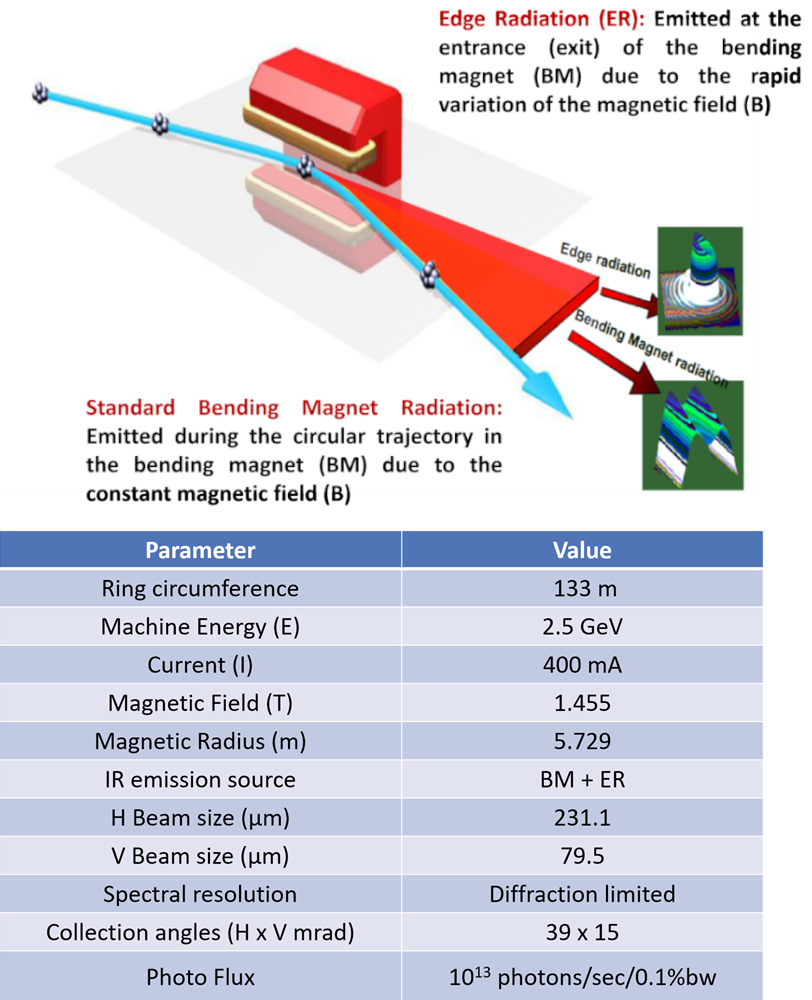
The SESAME BM02-IR beamline, operational since November 2018, is a cutting-edge facility serving the global Infrared scientific community with reliable capacity and advanced capabilities. The beamline was designed and built in collaboration with the French light source, SOLEIL. Dedicated for Synchrotron Radiation Fourier Transform Infrared Spectromicroscopy (SR-µFTIR), it uses a high-performance FTIR interferometer and harnesses infrared synchrotron radiation from both the bending magnet (BM) and edge radiation (ER) sources.
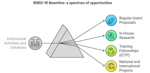
This vibrational technique, when coupled with synchrotron brilliance, delivers up to 1000× brighter signals than conventional thermal sources, with unique advantages in time structure, polarization, and broad wavelength coverage spanning the mid-IR (2.5–25 µm) and extending into the far-IR/THz region. The beamline provides powerful tools for non-destructive and spatially resolved chemical analysis. By integrating Fourier Transform Infrared (FTIR) spectrometry with advanced microscopy, the beamline enables scientists to identify and image IR-active vibrational modes at the diffraction-limited microscopic scale. In particular, it measures optically active molecular vibrations at the micro- and nano-scale, generating highly specific, spatially distributed chemical fingerprints. Diverse fields from material science and forensics, geology to mineralogy, food and agriculture, water and soil pollution and environmental studies, to pharma, biomedicine and life sciences, as well as cultural heritage, archaeology, palaeontology and art restoration.
With diffraction-limited spatial resolution and superior signal-to-noise ratios, the BM02-IR beamline opens new frontiers for discovery, offering scientists a powerful tool to explore the molecular world in unprecedented detail.
SESAME Joins INFN-CHNet with New Cultural Heritage Laboratory
In 2022, SESAME inaugurated a new laboratory dedicated to cultural heritage as part of the Cultural Heritage Network, CHNet, of the Italian National Institute for Nuclear Physics (INFN). This important milestone marked SESAME’s official inclusion in the network, reinforcing international collaboration. To support advanced research, INFN has equipped the BM02-IR beamline with a state-of-the-art endstation providing powerful tools for non-invasive analysis, enabling researchers to explore and safeguard cultural treasures. For more information, click here
Infrared (IR) spectromicroscopy is a non-destructive vibrational technique that has gained strong interest at synchrotron facilities worldwide. It combines the spatial resolution of a microscope with the high chemical sensitivity of an FTIR spectrometer, offering powerful insights into material composition.
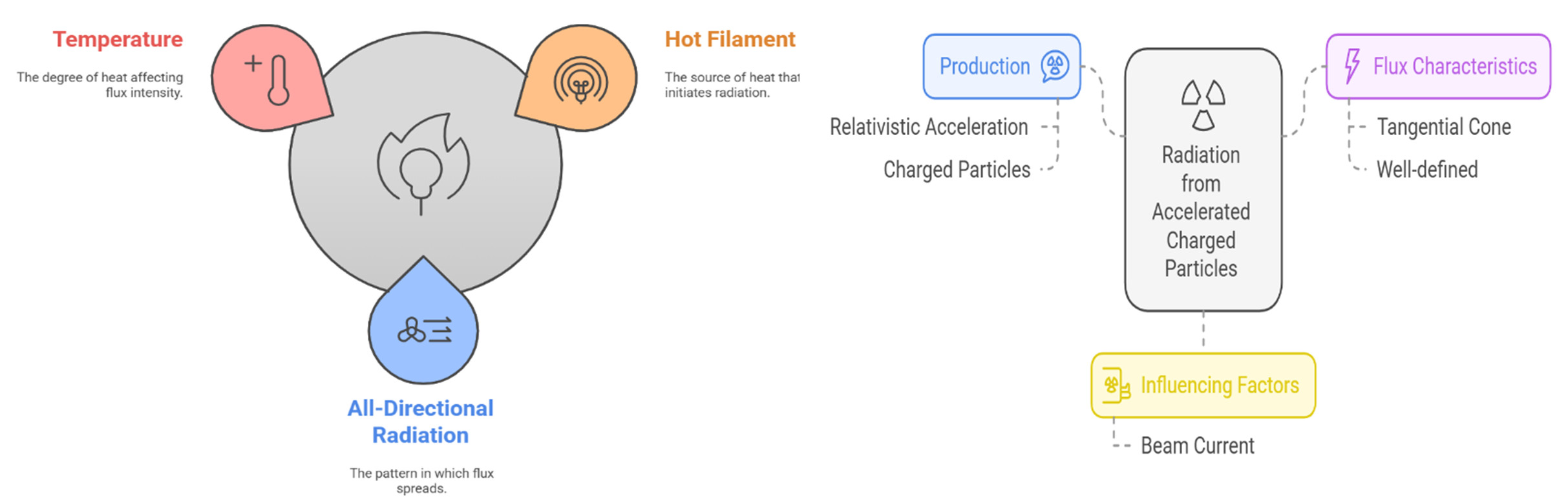
Synchrotron IR sources provide significant advantages over conventional Globar sources. Thanks to their brightness and brilliance, around 1000 times higher, they deliver a superior signal-to-noise ratio. The mid-infrared region (2.5–25 µm / 4000–400 cm⁻¹) is particularly informative at the microscopic scale, since most molecular groups exhibit vibrational energies in this range. Importantly, spatial resolution is determined not by aperture size but by the numerical aperture of the optical system and the wavelength of light, achieving diffraction-limited spot sizes of ~3–10 µm in confocal geometry.
Conventional sources typically use less than 1% of emitted light in diffraction-limit setups, whereas synchrotron sources are optimally matched, ensuring efficient use of radiation.
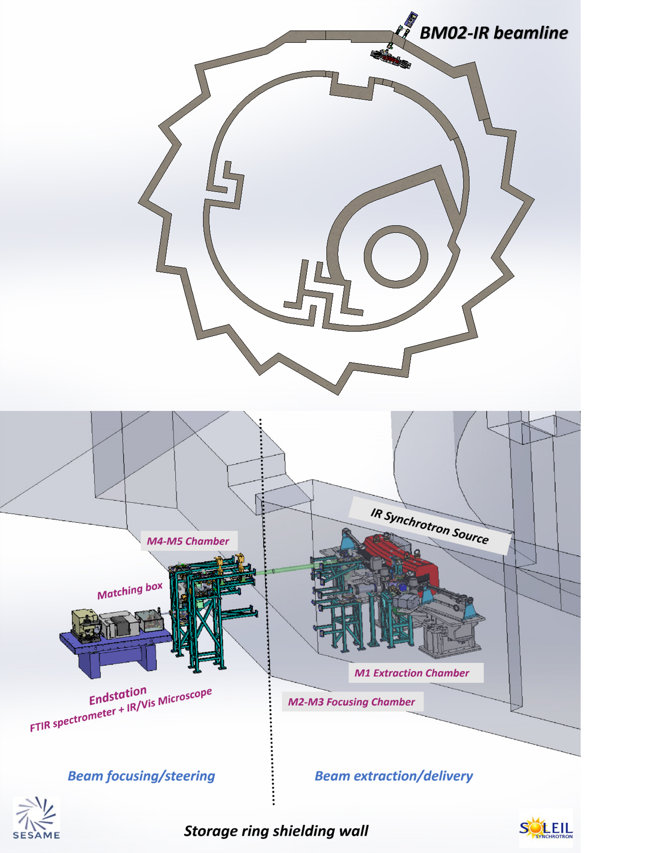
At SESAME, the BM02-IR beamline has been designed to cover both the mid-IR (2.5–25 µm) and far-IR (25–1000 µm) regions. With a maximum acceptance opening angle of 39 mrad (H) × 15 mrad (V), it harnesses infrared synchrotron emission from two radiation sources: the bending magnet’s constant field (BM) and edge radiation (ER). The optical system was carefully engineered to transport SRIR radiation to the experimental endstation with minimal aberrations, ensuring optimal beam delivery.
To achieve this, SESAME’s design was validated using advanced simulation software packages such as SRW and SPOT X, guaranteeing precision in beamline performance and reliability for cutting-edge research.
Dual-Beam Design and Future Scalability
The SESAME BM02-IR beamline has been purposefully engineered with scalability at its core. Its dual-beam design delivers radiation to the experimental floor, enabling the operation of two independent end stations without requiring any redesign of the original setup.
During the initial development phase, one end station will be made operational. As user demand grows and scientific applications diversify, a second end station can be seamlessly integrated into the system. This forward-looking design ensures SESAME can adapt to evolving research needs with minimal effort.
To support this flexibility, an easy mirror-swapping mechanism has been incorporated. This allows simultaneous operation of both modes or end stations, maximizing efficiency, adaptability, and productivity for a wide range of experiments.
Synchrotron beam Coupling with the FTIR Spectrometer
The coupling box, positioned between the final KBr window (beamline termination) and the FTIR spectrometer, reshapes the divergent rectangular beam into a more collimated form. This collimated beam enters the interferometer and can be adjusted along the X and Z axes, as well as tilted, to optimize alignment with the IR/Vis microscope.
Experimental endstations – Instrumentation and Setup
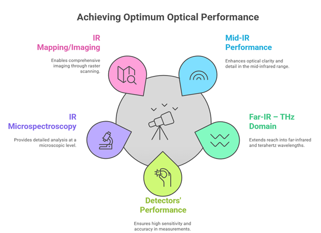
The BM02-IR beamline endstation has been designed for optimum performance across the mid- to far-IR range. A comprehensive suite of detectors, both single point and array, equip the endstation to fully cover the IR spectrum.
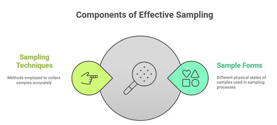
To meet the wide variety of scientific applications, the endstation offers a versatile range of sample environments and sampling methods. These are complemented by off-line services for sample preparation, handling, and advanced data analysis, ensuring researchers have the tools and support needed for high-quality investigations.
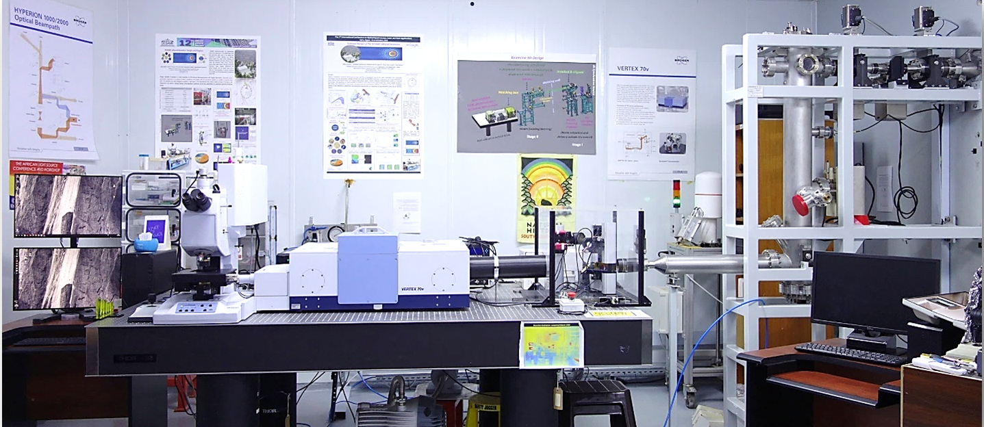
The SESAME BM02-IR beamline is currently equipped with state-of-the-art endstation of the Bruker© Vertex 70v FTIR FTIR spectrometer coupled to Hyperion 3000 IR-vis Microscope.
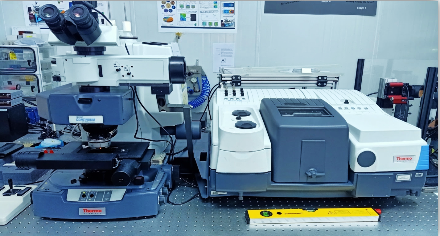
Additionally, the beamline is also giving access to a second offline endstation (Globar IR source) equipped with Thermo Scientific© 8700 FTIR spectrometer coupled to Nicolet Continuum XL IR-microscope.
BM D02
Offline-mode Endstation (Globar IR Source)
The microscope features Kohler Illumination for Reflection and Transmission, Brightfield, Darkfield and Polarized Light. In addition to DIC (Differential Interference Contrast) Optics; and Fluorescence illumination of:
-Wide Band Blue Fluorescence Cube 450-480nm,
-Wide Band Green Fluorescence Cube 510-550nm,
-Wide Band UV Fluorescence Cube 330-385nm.
DTGS
InGaAs
Sample
Specac Mini Pellet Press™ | FTIR transmission mode,
Specac Micro Compression Cell™ | FTIR transmission mode,
Specac DC-3 | Diamond Compression Cell™ | FTIR microscopy in transmission mode,
Specac Monolayer Grazing Angle Specular Reflectance Accessory™ | FTIR specular reflection mode,
Pike Technologies MIRacle™ cell | FTIR single reflection mode,
Thermo Fisher Slide-on 15x ATR Si Objective™
Manipulator or Sample stage
Sample Holders
Specac Mini Pellet Press™ | FTIR transmission mode,
Specac Micro Compression Cell™ | FTIR transmission mode,
Specac DC-3 | Diamond Compression Cell™ | FTIR microscopy in transmission mode,
Specac Monolayer Grazing Angle Specular Reflectance Accessory™ | FTIR specular reflection mode,
Pike Technologies MIRacle™ cell | FTIR single reflection mode,
Thermo Slide-on 15x ATR Si Objective™
Standard SR-IR Endstation
DTGS
InGaAs
Focal Plane Array (FPA)
Sample
Specac Mini Pellet Press™ | FTIR transmission mode,
Specac Micro Compression Cell™ | FTIR transmission mode,
Specac DC-3 | Diamond Compression Cell™ | FTIR microscopy in transmission mode,
Specac Monolayer Grazing Angle Specular Reflectance Accessory™ | FTIR specular reflection mode,
Pike Technologies MIRacle™ cell | FTIR single reflection mode,
Bruker 20x ATR Ge Objective™
Sample Holders
Specac Mini Pellet Press™ | FTIR transmission mode,
Specac Micro Compression Cell™ | FTIR transmission mode,
Specac DC-3 | Diamond Compression Cell™ | FTIR microscopy in transmission mode,
Specac Monolayer Grazing Angle Specular Reflectance Accessory™ | FTIR specular reflection mode,
Pike Technologies MIRacle™ cell | FTIR single reflection mode,
Bruker 20x ATR Ge Objective™
DTGS
- TE Cooled DLaTGS Detector with KBr Window (12,500-350 cm-1);
- DLaTGS Detector with Polyethylene Window (700-50 cm-1).
Detection
Focal Plane Array (FPA)
Detection
InGaAs
InGaAs detector for NIR (12.000-3.800 cm-1).
Detection
MCT
- 50um MCT-A Detector;
- MCT-B Detector.
Detection
Note: This input is synced with WayForLight.
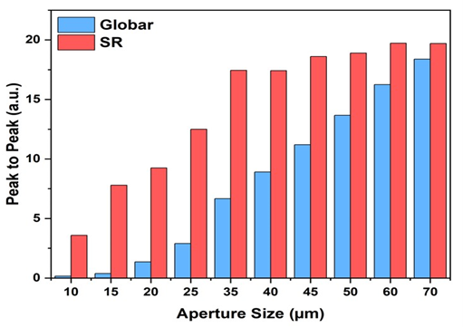
Peak-to-Peak interferogram values for both the SESAME BM02-IR beamline SR-IR (at 200 mA) and the Globar source at different aperture sizes. [Gihan Kamel et al. First IR beamline at SESAME, J. Synchrotron Rad. (2021), 28, 1927–1934]
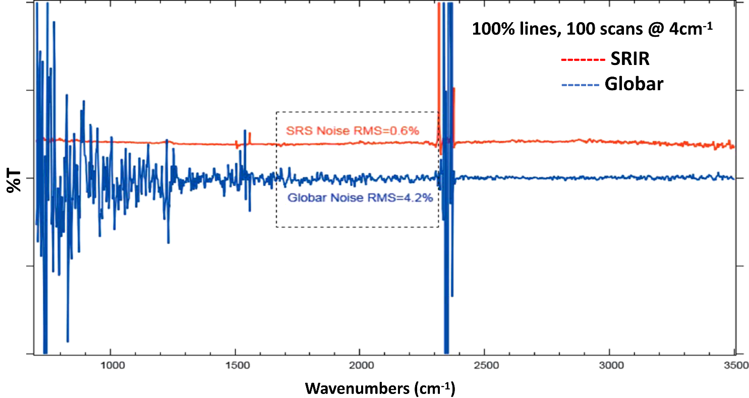
100% lines of SR-IR and Globar signals through a 20µmx20µm aperture demonstrating their signal-to-noise ratio. The SRS 100% line has been offset for clarity. [Gihan Kamel et al. First IR beamline at SESAME, J. Synchrotron Rad. (2021), 28, 1927–1934]
Molecular Spectroscopy and Vibrational Modes
Molecular spectroscopy uses electromagnetic radiation as a physical stimulus to probe the vibrational properties of molecules. When infrared (IR) radiation interacts with matter, it excites specific vibrational modes of chemical bonds, producing a spectrum that serves as a molecular fingerprint. The level of detail achieved depends on the instrumentation and the IR source whether conventional IR, synchrotron radiation, or IR lasers.
Key Vibrational Modes
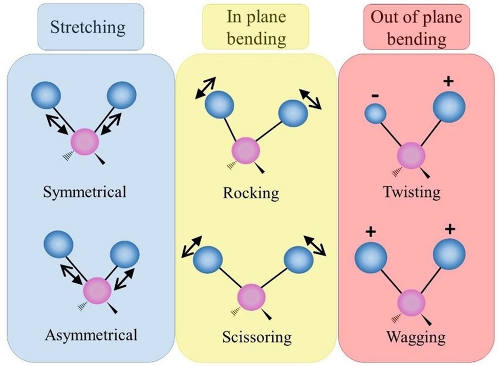
- Stretching vibrations (Symmetric and Asymmetric): involve the periodic lengthening and shortening of chemical bonds.
- Bending vibrations: Changes in bond angles rather than bond lengths.
- Scissoring: two atoms move toward and away from each other.
- Rocking: atoms move in the same direction, like a pendulum.
- Wagging: atoms move up and down out of the molecular plane.
- Twisting: atoms move in opposite directions out of the plane.
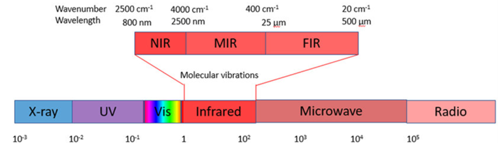
These vibrational modes occur in the mid-infrared region (2.5–25 µm / 4000–400 cm⁻¹), where most functional groups have characteristic absorption bands. This makes IR spectroscopy especially powerful for identifying molecular structures and functional groups.
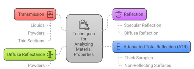
Infrared spectra can be collected using several complementary modes, each suited to specific sample types and research needs:
- Transmission mode: The simplest approach, requiring thin samples typically 5–30 µm in thickness. It is ideal for polymers, biological tissues, geological specimens, and organic materials.
- Reflection mode: Here, the IR beam penetrates the sample, reflects off the substrate, and passes back through the objective. This method is best for highly reflective or polished samples that cannot be sectioned thinly. Coating thicknesses usually range from 1-10 µm, and smooth, flat surfaces are essential to minimize scattering and background oscillations. In special cases, irregular surfaces can be pressed to align with the incident light and microscope stage.
- Attenuated Total Reflection (ATR): ATR probes the near-surface region of materials and is particularly useful for liquids, semi-solids, and pliable solids such as rubber and plastics. The sample is placed in contact with a high-refractive-index crystal (ZnSe, Ge, Si, or diamond), where total internal reflection generates an evanescent wave that penetrates the sample surface. This technique is effective for opaque or thick samples unsuitable for transmission.
- Grazing incidence FTIR: A specialized method for analyzing thin films on reflective substrates, often less than 1 µm thick. Measurements are taken at a controlled angle of incidence, using a Schwarzschild objective with a beam mask to transmit only grazing rays. This enhances sensitivity to ultra-thin coatings.
Modalities
FTIR spectroscopy
IR Microscopy: chemical mapping, chemical imaging.
Equipment available on site
Specac Omni-Cell™ | Demountable Liquid Cell for FTIR transmission mode
Specac Mini Pellet Press™ | FTIR transmission mode
Specac Micro Compression Cell™ | FTIR transmission mode
Specac DC-3 | Diamond Compression Cell™ | FTIR microscopy in transmission mode
Specac Monolayer Grazing Angle Specular Reflectance™ | FTIR specular reflection mode
Pike Technologies MIRacle™ cell | FTIR single reflection mode
Bruker 20x ATR Ge Objective™
Thermo Scientific Slide-on 15x ATR Si Objective™
Leica DMi1 inverted microscope™
Optika B-350 optical Stereomicroscope™
SLEE MEV + cryostat™
Shenyang Kejing SYJ-150 Precision Low Speed Diamond Saw™
Leco PX300 Dual Grinder-Polisher System™
Equipment that can be brought by the user
Temperature and pressure cells for in-situ measurements.
Possibility to connect external endstation
Thermo Scientific TOM Optical Module™ | Polarization-Modulation for:
(i) IRRAS: Infrared Reflection-Absorption Spectroscopy. (ii) VLD: Vibrational Linear Dichroism (iii) DIRLD: Dynamic Infrared Linear Dichroism.
- Control software type
- Beamline Control: EPICS© (EPICS - Experimental Physics and Industrial Control System)
- Beamline Standard endstation: OPUS® 8.5 (SP1) Build 8,7,10 Proprietary software
- (©Bruker Optik GmbH 2020).
- Beamline offline endstation: OMNIC® 9.2.41, OMNICMC® v 9.1.0, OMNIC Atlus® 9.1.24. (©Thermo Fisher Scientific Inc. 1992-2012)
- Data output type
Transmittance/Reflectance/Absorbance IR single spectra, interferograms, visible images, and hyperspectral images/chemical maps.
- Data output format
- Standard endstation: OPUS files as single spectra and maps
- Offline endstation: OMNIC files as single spectra and maps
- Software for data analysis
The BM02-IR beamline offers on-site and remote access to the following packages:
- OPUS® 8.5 (SP1) Build 8,7,10 (©Bruker Optik GmbH 2020),
- OMNIC® 9.2.41, OMNICMC® v 9.1.0, OMNIC Atlus® 9.1.24. (©Thermo Fisher Scientific Inc. 1992-2012),
- The Unscrambler X® v 10.4. (© CAMO Analytics),
- CytoSpec® v 1.3.02. (©2000-2020 Peter Lasch),
- PeakFit v4.12 (©Systat Software, USA),
- Quasar®.
2025 (6), 2024 (3), 2023 (2), 2022 (7), 2021 (6), 2020 (2), 2017 (2), 2016 (3), 2012 (1), All (32), Thesis (1)
2025
- Revealing sassanid dyeing practices through synchrotron FTIR,
Journal: Scientific Reports,
Vol. 15 - 1, pp. 41318 (2025)
M.H. Dehkordi, G. Kamel, A.S.H. Rozatian, F.S. Madani, S. Noohi, A. Refaat, J. Nokandeh, M. Emami
doi: 10.1038/s41598-025-25106-0 - Synchrotron FTIR microspectroscopy for quantitative assessment of collagen mineralization: a comparative analytical study,
Journal: Analytical and Bioanalytical Chemistry (2025)
K. Hacıosmanoglu, A. Belet, G. Kamel, M. Kazanci
doi: 10.1007/s00216-025-06204-4 - Transit Phases and Beta Amyloid Aggregation/Clearance in Al-Induced Alzheimer's Disease in Rat Brain Hippocampus: Synchrotron Fourier Transform Infrared Microspectroscopy (SFTIRM) Study,
Journal: The Journal of Neurology & Neuropsychiatry,
Vol. 2 - 1, pp. 30-43 (2025)
S. Khalil, W. ElHotaby, G. Ahmed, G. Kamel, H. Sherif, L. Abbas
doi: 10.X - Development and characterization of crosslinked collagen biomaterial inks for 3D bioprinting applications,
Journal: Biomedical Materials,
Vol. 20 (2025)
A. Belet, K. Selcuk Haciosmanoglu, E. Atas, U. Demir, G. Kamel, M. Kazanci
doi: 10.1088/1748-605X/ae142e - Confirmation of L-phenylalanine's toxic fibrillary formation and its modulation by D-phenylalanine at different ratios and pH values by using synchrotron FTIR,
Journal: Spectrochimica Acta - Part A: Molecular and Biomolecular Spectroscopy,
Vol. 333 (2025)
S. K. Hacıosmanoglu, M. G. Akkurt, E. Atas, G. Kamel, S. Uyaver, M. Kazanci
doi: 10.1016/j.saa.2025.125891 - Insights into the anticancer and anti-inflammatory activities of curcumin-loaded quercetin nanoparticles: in vitro bioassays coupled with synchrotron infrared microspectroscopy,
Journal: Mater. Adv. (2025)
S Sunoqrot, S Abusulieh, LA Dahabiyeh
doi: 10.1039/D4MA01202J
- Biochemical analysis of Hyalomma dromedarii salivary glands and gut tissues using SR-FTIR micro-spectroscopy,
Journal: Scientific Reports,
Vol. 14 - 1, pp. 8515 (2024)
S.H.M. Hendawy, H.F. Alzan, H.S.M. Abdel-Ghany, C.E. Suarez, G. Kamel
doi: 10.1038/s41598-024-59165-6 - Green plastics: Direct production from grocery wastes to bioplastics and structural characterization by using synchrotron FTIR,
Journal: Spectrochimica Acta Part A: Molecular and Biomolecular Spectroscopy,
Vol. 323, pp. 124919 (2024)
O Aras, G Kamel, M Kazanci
doi: 10.1016/j.saa.2024.124919 - A Comprehensive Study on How Different Mineralization Processes Affect Native Structure of Wetspun Collagen Fibers and Mineral Quality by Using S(Ftir),
Journal: Elsevier SSRN (Social Science Research Network) (2024)
K. Selcuk Haciosmanoglu, A. Belet, G. Kamel, M. Kazanci
doi: 10.2139/ssrn.4775064
- Synchrotron Fourier Transform Infrared Microspectroscopy (Sftirm) Analysis of Beta Amyloid Aggregation/Clearance in Al-Induced Alzheimer'S' Disease in Rat Brain Hippocampal Tissue,
Journal: Elsevier SSRN (Social Science Research Network) (2023)
S. K. H. Khalil, W. El hotaby, G. Abdel-Raouf Ahmed, G. Kamel, H. H. A. Sherif, L. Abbas
doi: 10.2139/ssrn.4560767 - IR microspectroscopic investigation of the interaction of some losartan salts with human stratum corneum protein and its effect on losartan transdermal permeation,
Journal: PLOS ONE,
Vol. 18 - 6, pp. e0287267 (2023)
R.S.H. Mansour, A.Y. Al Khawaja, I.I. Hamdan, E.A. Khalil
doi: 10.1371/journal.pone.0287267
- Synchrotron radiation infrared microspectroscopy: Insights on biomedicine,
Journal: Applied Spectroscopy Reviews, pp. 1-20 (2022)
A. Refaat, G. Kamel
doi: 10.1080/05704928.2022.2052308 - Synchrotron Fourier-transform infrared microspectroscopy: Characterization of tumor infiltrating monocytes stimulated by the secretome of inflammatory and non-inflammatory breast cancer cells,
Conference: AACR Annual Meeting 2022,
New Orleans (US) 18/11/2021 (2022)
A. El-Sharkawy, G. Kamel, H. Mohamed, M. El-Shinawi, M. Mohamed, N. El-Husseiny
doi: 10.1158/1538-7445.AM2022-6132 - Plasma drop and thin-film revealed distinguished molecular structure in pre-eclampsia: An investigation using synchrotron Fourier-transform infrared microspectroscopy,
Journal: Journal of Pharmaceutical and Biomedical Analysis,
Vol. 220, pp. 114981 (2022)
L.A. Dahabiyeh, R.S.H. Mansour, W. Darwish, S.S. Saleh, G. Kamel
doi: 10.1016/j.jpba.2022.114981 - Synchrotron Fourier-Transform Infrared Microspectroscopy: Characterization of in vitro polarized tumor-associated macrophages stimulated by the secretome of inflammatory and non-inflammatory breast cancer cells,
Journal: Biochimica et Biophysica Acta - Molecular Cell Research (2022)
H. Mohamed, G. Kamel, N. El-Husseiny, A. El-Sharkawy, A. El-Sherif, M. El-Shinawi, M. Mohamed
doi: 10.1016/j.bbamcr.2022.119367 - Synchrotron-Radiation-Based Fourier Transform Infrared Microspectroscopy as a Tool for the Differentiation between Staphylococcal Small Colony Variants,
Journal: Antibiotics,
Vol. 11 - 11 (2022)
AG Al-Bakri, LA Dahabiyeh, E Khalil, D Jaber, G Kamel, N Schleimer, C Kohler, K Becker
doi: 10.3390/antibiotics11111607 - Investigating the molecular structure of plasma in type 2 diabetes mellitus and diabetic nephropathy by synchrotron Fourier-transform infrared microspectroscopy,
Journal: Spectrochimica Acta Part A: Molecular and Biomolecular Spectroscopy,
Vol. 264, pp. 120259 (2022)
R. Nimer, G. Kamel, MA. Obeidat, LA. Dahabiyeh
doi: 10.1016/j.saa.2021.120259 - Synchrotron Radiation Fourier Transform Infrared (SR-FTIR) spectroscopy in exploring ancient human hair from Roman period Juliopolis: Preservation status and alterations of organic compounds,
Journal: Spectrochimica Acta Part A: Molecular and Biomolecular Spectroscopy, pp. 121026 (2022)
K.O. Lorentz, G. Kamel, S.AM. Lemmers, Y. Miyauchi, E. Cubukcu, A. Alpagut, AM. Buyukkarakaya
doi: 10.1016/j.saa.2022.121026
- Characterization of Insulin Mucoadhesive Buccal Films: Spectroscopic Analysis and In Vivo Evaluation,
Journal: Symmetry,
Vol. 13 - 1, pp. 1-17 (2021)
M Diab, A Sallam, I Hamdan, R Mansour, R Hussain, G Siligardi, N Qinna, E Khalil
doi: 10.3390/sym13010088 - Investigation of ancient teeth using Raman spectroscopy and synchrotron radiation Fourier-transform infrared (SR-muFTIR): mapping and novel method of dating
,
Journal: Digest Journal of Nanomaterials and Biostructures,
Vol. 16, pp. 713-724 (2021)
W. Sekhaneh, Y. Akkam, G. Kamel, A. Drabee, J. Popp
doi: 10.15251/djnb.2021.162.713 - Transcriptional modulations induced by proton irradiation in mice skin in function of adsorbed dose and distance,
Journal: Journal of Radiation Research and Applied Sciences,
Vol. 14 - 1, pp. 260-270 (2021)
V. Licursi, W. Wang, E. Di Nisio, F.P. Cammarata, R. Acquaviva, G. Russo, L. Manti, M. Cestelli Guidi, E. Fratini, G. Kamel, R. Amendola, P. Pisciotta, R. Negri
doi: 10.1080/16878507.2021.1949675 - Mummified Embalmed Head Skin: SR-FTIR Microspectroscopic Exploration,
Journal: Spectrochimica Acta Part A: Molecular and Biomolecular Spectroscopy, pp. 120073 (2021)
D. Moissidou, H. Derricott, G. Kamel
doi: 10.1016/j.saa.2021.120073 - The first infrared beamline at the Middle East SESAME synchrotron facility.,
Journal: Journal of Synchrotron Radiation,
Vol. 28 - 6 (2021)
G. Kamel, S. Lefrancois, T. Moreno, M. Al-Najdawi, Y. Momani, A. Abbadi, G. Paolucci, P. Dumas
doi: 10.1107/S1600577521008778 - Synchrotron Fourier transform infrared microspectroscopy (sFTIRM) analysis of unfolding behavior of electrospun collagen nanofibers,
Journal: Spectrochimica Acta - Part A: Molecular and Biomolecular Spectroscopy,
Vol. 251 (2021)
M. Kazanci, K. Selcuk Haciosmanoglu, G. Kamel
doi: 10.1016/j.saa.2020.119420
- Investigating the molecular structure of placenta and plasma in pre-eclampsia by infrared microspectroscopy,
Journal: Journal of Pharmaceutical and Biomedical Analysis,
Vol. 184, pp. 113186 (2020)
L.A. Dahabiyeh, R.S.H. Mansour, S.S. Saleh, G. Kamel
doi: 10.1016/j.jpba.2020.113186 - Synchrotron Fourier transform infrared microspectroscopy (sFTIRM) analysis of Al-induced Alzheimer's disease in rat brain cortical tissue,
Journal: Spectrochimica Acta Part A: Molecular and Biomolecular Spectroscopy,
Vol. 239, pp. 118421 (2020)
G.A. Ahmed, W. El Hotaby, L. Abbas, H.H.A. Sherif, G. Kamel, S.KH. Khalil
doi: 10.1016/j.saa.2020.118421
- EMIRA: The Infrared Synchrotron Radiation Beamline at SESAME,
Journal: Synchrotron Radiation News,
Vol. 30 - 4, pp. 8-10 (2017)
G. Kamel, S. Lefrancois, M. Al-Najdawi, T. Abu-Hanieh, I. Saleh, Y. Momani, P. Dumas
doi: 10.1080/08940886.2017.1338415 - Elucidation of penetration enhancement mechanism of Emu oil using FTIR microspectroscopy at EMIRA laboratory of SESAME synchrotron,
Journal: Spectrochimica Acta Part A: Molecular and Biomolecular Spectroscopy,
Vol. 185, pp. 1-10 (2017)
R.S.H. Mansour, A.A. Sallam, I.I. Hamdan, E.A. Khalil, I. Yousef
doi: 10.1016/j.saa.2017.05.026
- Optical and Micro-FTIR mapping: A new approach for structural evaluation of V2O5-lithium fluoroborate glasses,
Journal: Materials Design,
Vol. 89, pp. 568-572 (2016)
A.M. Abdelghany, H.A. ElBatal
doi: 10.1016/j.matdes.2015.09.159 - Monosialoganglioside-GM1 triggers binding of the amyloid-protein salmon calcitonin to a Langmuir membrane model mimicking the occurrence of lipid-rafts,
Journal: Biochemistry and Biophysics Reports,
Vol. 8, pp. 365-375 (2016)
M. Diociaiuti, C. Giordani, G. Kamel, F. Brasili, S. Sennato, C. Bombelli, K. Meneses, M. Giraldo, F. Bordi
doi: 10.1016/j.bbrep.2016.10.005 - Study of the biochemical effects induced by X-ray irradiations in combination with gadolinium nanoparticles in F98 glioma cells: first FTIR studies at the Emira laboratory of the SESAME synchrotron,
Journal: Analyst,
Vol. 141 - 7, pp. 2238-2249 (2016)
I. Yousef, O. Seksek, S. Gil, Y. Prezado, J. Sule-Suso, I. Martinez-Rovira
doi: 10.1039/C5AN02378E
- Simulation and design of an infrared beamline for SESAME (Synchrotron-Light for Experimental Science and Applications in the Middle East),
Journal: Nuclear Instruments and Methods in Physics Research Section A: Accelerators, Spectrometers, Detectors and Associated Equipment,
Vol. 673, pp. 73-81 (2012)
I. Yousef, S. Lefrancois, T. Moreno, H. Hoorani, F. Makahleh, A. Nadji, P. Dumas
doi: 10.1016/j.nima.2011.12.012



Gihan KAMEL
IR Beamline Principal Scientist and Team Supervisor
Email: gihan.kamel@sesame.org.jo
Work Tel: +962 5 351 1348 (Ext. 240)
Yuko MIYAUCHI
JSPS Overseas Research Fellow
Email: yuko.miyauchi@sesame.org.jo
Work Tel: +962 5 351 1348 (Ext. 275)