
In an innovative study combining nanotechnology and synchrotron science, researchers from Al-Zaytoonah University and the University of Jordan, in collaboration with SESAME’s IR Beamline Scientist, Gihan Kamel, have developed a novel nanoformulation that significantly enhances the anticancer and anti-inflammatory potential of curcumin (CUR).
The study, published in the Royal Society of Chemistry's journal Materials Advances, focuses on leveraging quercetin-based nanoparticles to overcome CUR’s poor solubility and limited bioavailability—key barriers in its clinical use.
To gain deeper biochemical insights into the cellular response, researchers employed Synchrotron Fourier-Transform Infrared Microspectroscopy (SR-FTIRµ) at SESAME’s BM02-IR beamline. This advanced bioanalytical technique revealed notable biochemical changes in the lipid and protein compositions of treated cells, offering a molecular-level understanding of the nano-formulation’s therapeutic action. The integration of SR-FTIRµ helped in validating the nanoparticle’s multimodal bioactivity, reinforcing the relevance of synchrotron methods in drug development and biomedicine. (Fig.1)
The full study is available at: https://doi.org/10.1039/D4MA01202J
S.Sunoqrot, S. Abusulieha, L. A. Dahabiyeh
Materials Advances, 2025,6, 1971-1987.
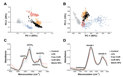
Fig. 1 SR-FTIRµ measurements of the lipid and protein-carbonyl regions. PCA-score plots of the (A) lipid and (B) protein-carbonyl regions. Control: black dots; LPS-stimulated cells: orange dots; free CUR: blue dots; CUR NPs: pink dots; blank NPs: grey dots; unit vector normalized average raw spectra of the (C) lipid and (D) protein-carbonyl regions.